The brain's cerebral hemispheres
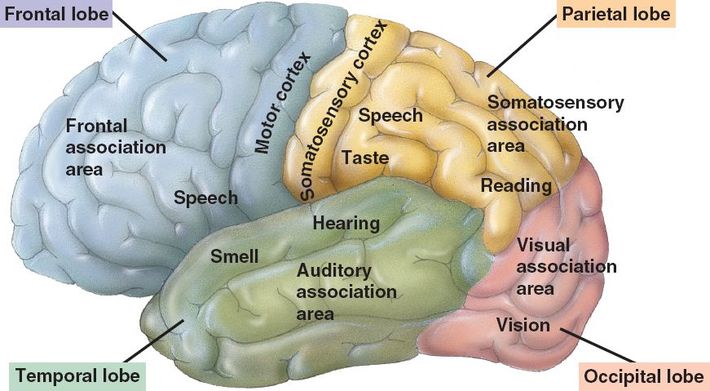
The cerebral hemispheres form the uppermost portion of the brain and between the two of them, they account for approximately 83% of the total mass of the brain. They are the most obvious portions of an intact brain. The paired cerebral hemispheres are like a dome that covers and obscures that diencephalon and the top portion of the brain stem. Almost the entire surface of the cerebral hemispheres are marked by gyri, which are elevated ridges of tissue, separated by shallow and narrow grooves called sulci. Fissures are deeper grooves that separate large regions of the brain. The more prominent gyri and sulci are very similar in every person and are very important as anatomical landmarks. There is a median longitudinal fissure that separates the two cerebral hemispheres and the transverse cerebral fissure that separates the two cerebral hemispheres from the cerebellum, located beneath it. Multiple sulci divide each of the cerebral hemispheres into five different lobes: frontal, parietal, temporal, occipital, and the insula. Each are named for the bones that cover them except for the insula. The central sulcus,located within the frontal brain, separates the from the frontal lobe from the parietal lobe. Lying alongside the central sulcus are the precentral gyrus (located anteriorly) and the postcentral gyrus (located posteriorly). Furthest back is that occipital lobe which is separated from the parietal lobe by the parieto-occipital sulcus. The temporal lobe, which is rather flap-like, is separated from the parietal and frontal lobes by the deep lateral sulcus. The fifth lobe of the cerebral hemisphere is the insula, which is covered by portions of the temporal, parietal, and frontal lobes and buried within the lateral sulcus, part of its floor.
The cerebral hemispheres are snugly fit into the skull. The frontal lobes are set in the anterior cranial fossa. The middle cranial fossa support the anterior parts of the temporal lobes. The cerebellum and brain stem are housed within the posterior cranial fossa, and the occipital lobes are found superior to that fossa.
The cerebral hemispheres are snugly fit into the skull. The frontal lobes are set in the anterior cranial fossa. The middle cranial fossa support the anterior parts of the temporal lobes. The cerebellum and brain stem are housed within the posterior cranial fossa, and the occipital lobes are found superior to that fossa.
cerebral cortex
The conscious mind is found in the cerebral cortex. Self-awareness, sensation, communication, memory, understanding, and voluntary motions all come from the cerebral cortex. It is made of gray matter: neuron cell bodies, dendrites, associated glia and blood vessels, but no fiber tracts. It has six layers composed of billions of neurons each. Accounting for roughly 40% of the brain's total mass, it is only about 1/8 inch thick and has a surface area more than 3 times its appearance due to its many convolutions. There are four generalizations that can be made about the entire cerebral cortex:
Motor areas:
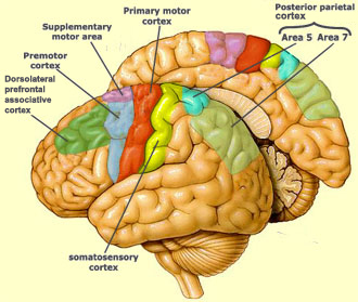
Motor areas control voluntary movement and lie in the posterior part of the frontal lobes. There are four major parts: primary motor cortex, premotor cortex, Broca's area, and the frontal eye field.
Primary motor cortex: Located in the precentral gyrus of the frontal lobe of each hemisphere, the primary motor cortex contains large pyramidal cells, a specialized form of neuron, which allow conscious control over the movements of skeletal muscles. Their axons form long "pyramidal (corticospinal) tracts" that extend to the spinal cord. An interesting note is that all the pyramidal cells controlling a specific part of the body are found grouped together, forming a "map" of the body through a phenomenon known as somatotopy. The body is represented upside down in this map; that is, the head is connected with the inferolateral part of the precentral gyrus, and the toes with the superomedial end. Most neurons in these gyri control muscles in body areas having the most precise motor control, as well as exhibiting contralateral control (the left primary motor gyrus controls the right side of the body and vice versa). It is noteworthy that, while these gyri control specifc parts of the body, there is not a one-to-one relationship between neurons and muscles. Instead, neurons coordinate with numerous muscles so as to create movements in useful ways (moving the arm requires multiple muscles all working in tandem).
Premotor cortex: Located just anterior to the precentral gyrus, the premotor cortex controls learned motor skills of a repetitious or patterned nature, like typing or playing a musical instrument. It specializes in coordinating the movement of several muscle groups either simultaneously or sequentially, mainly by sending activation impulses to the primary motor cortex. The premotor cortex also seems to be involved in planning movements and voluntary actions that depend on sensory feedback.
Broca's area: Located anterior to the inferior region of the premotor area, the Broca's area is quite unique in that it is considered to be present in one hemisphere only (typically the left). It directs the muscles involved in speech production and has been shown to activate upon planning to speak or planning of various other voluntary motor activities.
Frontal eye field: Located partially in and anterior to the premotor cortex and superior to Broca's area, this region controls voluntary movements of the eyes.
- It contains three kinds of functional areas: motor areas, sensory areas, and association areas.
- Each hemisphere is chiefly concerned with the sensory and motor functions of the opposite side of the body.
- Although largely symmetrical in structure, the two hemispheres are not entirely equal in function. Instead, there is a specialization of cortical functions.
- No functional area of the cortex acts alone, and conscious behavior involves the entire cortex in one way or another.
Motor areas:
Motor areas control voluntary movement and lie in the posterior part of the frontal lobes. There are four major parts: primary motor cortex, premotor cortex, Broca's area, and the frontal eye field.
Primary motor cortex: Located in the precentral gyrus of the frontal lobe of each hemisphere, the primary motor cortex contains large pyramidal cells, a specialized form of neuron, which allow conscious control over the movements of skeletal muscles. Their axons form long "pyramidal (corticospinal) tracts" that extend to the spinal cord. An interesting note is that all the pyramidal cells controlling a specific part of the body are found grouped together, forming a "map" of the body through a phenomenon known as somatotopy. The body is represented upside down in this map; that is, the head is connected with the inferolateral part of the precentral gyrus, and the toes with the superomedial end. Most neurons in these gyri control muscles in body areas having the most precise motor control, as well as exhibiting contralateral control (the left primary motor gyrus controls the right side of the body and vice versa). It is noteworthy that, while these gyri control specifc parts of the body, there is not a one-to-one relationship between neurons and muscles. Instead, neurons coordinate with numerous muscles so as to create movements in useful ways (moving the arm requires multiple muscles all working in tandem).
Premotor cortex: Located just anterior to the precentral gyrus, the premotor cortex controls learned motor skills of a repetitious or patterned nature, like typing or playing a musical instrument. It specializes in coordinating the movement of several muscle groups either simultaneously or sequentially, mainly by sending activation impulses to the primary motor cortex. The premotor cortex also seems to be involved in planning movements and voluntary actions that depend on sensory feedback.
Broca's area: Located anterior to the inferior region of the premotor area, the Broca's area is quite unique in that it is considered to be present in one hemisphere only (typically the left). It directs the muscles involved in speech production and has been shown to activate upon planning to speak or planning of various other voluntary motor activities.
Frontal eye field: Located partially in and anterior to the premotor cortex and superior to Broca's area, this region controls voluntary movements of the eyes.
Sensory areas:
Sensory areas are concerned with conscious awareness of sensation and are found in the parietal, insular, temporal, and occipital lobes.
Primary somatosensory cortex: Located in the postcentral gyrus of the parietal lobe, just posterior to the primary motor cortex. Using sensory receptors in the skin and proprioceptors (position sense receptors) in the skeletal muscles, joints, and tendons, this part of the brain uses spatial discrimination to determine the body region being stimulated. Just like in the primary motor cortex, the body is represented upside down and contralaterally.
Somatosensory association cortex: Located just posterior to the primary somatosensory cortex, the two are highly connected. Primarily used to use data from the primary somatosensory cortex (temperature, pressure, etc.) to create an understanding of an object being felt.
Visual areas:
Primary visual (striate) cortex: Visible on the extreme posterior tip of the occipital lobe, but most of it is deep within the calcarine sulcus in the medial aspect of the occipital lobe. It receives visual information from the retina of the eye and is the largest of all cortical sensory areas. Just like on the somatosensory cortex, there is a contralateral map of the visual space on the primary visual cortex.
Visual association area: Surrounds the primary visual cortex and covers much of the occipital lobe. This area is responsible for using past experiences to make sense of the data from the primary visual cortex and allow us to recognize a person, object, etc. When processing information, there are two identifiable "streams" of visual data: one atop the brain responsible for evaluating spatial relationships and object location, and one lower in the brain responsible for identifying objects.
Auditory areas:
Primary auditory cortex: Located in the superior margin of the temporal lobe abutting the lateral sulcus. Auditory impulses from the inner ear are interpreted by this region as pitch, loudness, and location.
Auditory association area:
Located more posteriorly, the auditory association area interprets the sound stimulus as a voice, music, noise, etc. It is believed that memories of sounds from the past are stored here for reference.
Olfactory cortex:
Located on the medial aspect of the temporal lobe in a small region called the piriform lobe. Smell receptors in the superior nasal cavities send impulses along olfactory tracts which eventually reach the olfactory cortex, allowing us to smell different odors. The olfactory cortex is part of the rhinencephalon, a primitive portion of the brain made up of all parts of the cerebrum that receive olfactory signals: the orbitofrontal cortex, the uncus and associated regions on the temporal lobe, and the olfactory tracts and bulbs that extend to the nose. Most of these areas have been repurposed for dealing with emotions and memory in the limbic system. Only the olfactory bulbs and tracts and the olfactory cortex remain devoted to smell.
Gustatory cortex: Located in the insula just deep to the temporal lobe, it is responsible for the perception of taste stimuli.
Visceral sensory area: Cortex of the insula just posterior to the gustatory cortex. It interprets conscious visceral sensations (full bladder, upset stomach, need for air, etc.)
Vestibular cortex: Most likely located in the posterior part of the insula and adjacent to the parietal cortex, this region of the cortex is responsible for conscious awareness of balance and the position of the head in space.
Multimodal association areas: Areas of the cortex not tied to a specific sense, but receiving input from multiple senses and sending outputs to multiple areas. Information is typically sent to one of these areas after it has passed through a sensory association area. In the multimodal association area, the information is given meaning, stored in memory, connected with previous knowledge/ experience, and action is decided upon. It is believed that the multimodal association cortex is where sensations, thoughts, and emotions become consciousness.
Anterior association area: Located in the frontal lobe, the anterior association area, or prefrontal cortex, is the most complicated cortical region. Complex learning abilities, intellect, recall, and personality all highly involve this area. It is also vital to the production of abstract ideas, judgment, reasoning, persistence, and planning because it contains working memory.
Posterior association area: Encompasses parts of the temporal, parietal, and occipital lobes. The posterior association area is important in recognizing patters and faces, as well as localizing us and our surroundings in space and determining the area of space occupied by one's own body. It takes multiple sensory inputs and binds them into a coherent whole. Many parts of this area also become involved with understanding written and spoken language.
Limbic association area: Includes the cingulate gyrus, parahippocampal gyrus, and hippocampus. The limbic association area is part of the limbic system and provides the emotional impact that makes a scene or event important. The hippocampus establishes memories that allow us to remember the incident.
Lateralization of cortical functioning: While physically near-identical, each brain hemisphere has unique abilities not shared by its partner, a phenomenon called lateralization. One hemisphere or the other is dominant when performing various tasks, but the term cerebral dominance refers to the hemisphere that is dominant for language. Generally the left hemisphere is dominant for language, math, and logic, while the right is dominant in visual-spatial skills, intuition, emotion, art, and music. An important aspect of the brain is its complete functional integration, meaning that neither side of the brain can be superior at everything. This integration is made possible by the hemispheres having near instantaneous communication via connection fiber tracts.
Cerebral white matter
The second basic region of the brain is the cerebral white matter, which is responsible for communication between cerebral areas and between the cerebral cortex and lower CNS centers. White matter consists primarily of myelinated fibers bundled into large tracts, which can be classified as commissural, association, or projection.
Commissures: Composed of commissural fibers, these allow the two halves of the brain to function as a coordinated whole by connecting corresponding gray areas of the two hemispheres. The largest of these is the corpus callosum, which is superior to the lateral ventricles, deep within the longitudinal fissure. Two others are the anterior commissures and posterior commissures.
Association fibers: Association fibers connect different parts of the same hemisphere. Short ones connect adjacent gyri. Long ones are bundled into tracts and connect different cortical lobes.
Projection fibers: Connect the cerebral cortex to lower brain. Sensory input comes in through these fibers, and motor output leaves through them, making them the connection between the brain and the rest of the nervous system. Unlike commissural and association fibers, however, they run vertically, not horizontally. The projection fibers at the top of the brain stem form the internal capsule, a compact band that passes between the thalamus and some of the basal nuclei. After that, the fibers radiate out through the white matter to the cortex in an arrangement known as the corona radiata.
basal nuclei
Basal nuclei are found deep within the cerebral white matter. They are formed primarily by the caudate nucleus putamen and globus pallidus. The putamen and globus pallidus are create a mass on the lateral side of the internal capsule called the lentiform nucleus. The lentiform and caudate nuclei are collectively referred to as the corpus striatum due to their striped appearance from the fibers of the internal capsule. The basal nuclei receive input from the entire cerebral cortex and output to the premotor and prefrontal cortices. It is believed that they are involved in attention and cognition as well as starting, stopping, and monitoring the intensity of slow, stereotyped movements by the cortex, such as arm-swinging when walking.