Gross anatomy and protection of the spinal cord
The spinal cord runs from the opening in the base of the skull to the level of the first or second lumbar vertebrae, located just below the ribs. The spinal cord is forty-two centimeters long and three-quarters on an inch thick. It appears as a thick, white cord providing a two-way pathway for conduction to and from the brain. The spinal cord is also a major reflex center, and spinal reflexes are both initiated and completed at the level of the spinal cord.
Similar to the brain, the spinal cord is protected by bone, meninges, and cerebrospinal fluid. The spinal dura matter is single layered and not attached to the bony walls of the vertebral column. Between the vertebrae and the spinal dura matter is an epidural space filled with a soft padding of fat and a network of veins. Cerebrospinal fluid fills the subarachnoid space between the arachnoid and pia mater meninges. Beneath that, the dural and arachnoid membranes extend well below the end of the spinal cord, since the spinal cord usually terminates around the first or second lumbar vertebrae. Because of this, the subarachnoid space within the meningeal sac, which is below the that point, provides a perfect spot for removing cerebrospinal fluid for testing. This procedure is referred to as a lumbar puncture or tap. There is little to no danger of damaging the spinal cord or nerves because the cord is absent there and the nerve roots drift away from where the needle is inserted.
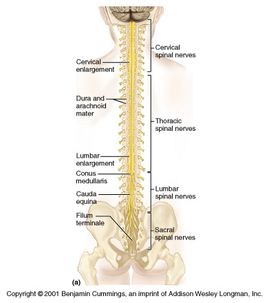
The spinal cord terminates in a tapering cone-shaped structure called the conus medullaris. There is a fibrous extension of the spinal cord called the filum terminale that extends into the coccyx, where it anchors the spinal cord so it is not jostled by body movements. It is further secured to the dura mater meninx throughout its length by saw-toothed shelves of pia mater called denticulate ligaments.
31 pairs of spinal nerves attach to the cord by paired roots, where each nerve exits the vertebral column by passing superior to its corresponding vertebra via the intervertebral foramen. Across its length, the spinal cord is about as large around as a thumb, but it has two major enlargements in the cervical and lumbosacral regions, where the nerves serving the upper and lower limbs arise. These enlargements are called the cervical and lumbar enlargements. Because the spinal cord does not reach the end of the vertebral column, the lumbar and sacral spinal nerve roots angle sharply downward and travel inferiorly through the vertebral canal for some distance before reaching their intervertebral foramina. This collection of nerve roots is called the cauda equina.
Similar to the brain, the spinal cord is protected by bone, meninges, and cerebrospinal fluid. The spinal dura matter is single layered and not attached to the bony walls of the vertebral column. Between the vertebrae and the spinal dura matter is an epidural space filled with a soft padding of fat and a network of veins. Cerebrospinal fluid fills the subarachnoid space between the arachnoid and pia mater meninges. Beneath that, the dural and arachnoid membranes extend well below the end of the spinal cord, since the spinal cord usually terminates around the first or second lumbar vertebrae. Because of this, the subarachnoid space within the meningeal sac, which is below the that point, provides a perfect spot for removing cerebrospinal fluid for testing. This procedure is referred to as a lumbar puncture or tap. There is little to no danger of damaging the spinal cord or nerves because the cord is absent there and the nerve roots drift away from where the needle is inserted.
The spinal cord terminates in a tapering cone-shaped structure called the conus medullaris. There is a fibrous extension of the spinal cord called the filum terminale that extends into the coccyx, where it anchors the spinal cord so it is not jostled by body movements. It is further secured to the dura mater meninx throughout its length by saw-toothed shelves of pia mater called denticulate ligaments.
31 pairs of spinal nerves attach to the cord by paired roots, where each nerve exits the vertebral column by passing superior to its corresponding vertebra via the intervertebral foramen. Across its length, the spinal cord is about as large around as a thumb, but it has two major enlargements in the cervical and lumbosacral regions, where the nerves serving the upper and lower limbs arise. These enlargements are called the cervical and lumbar enlargements. Because the spinal cord does not reach the end of the vertebral column, the lumbar and sacral spinal nerve roots angle sharply downward and travel inferiorly through the vertebral canal for some distance before reaching their intervertebral foramina. This collection of nerve roots is called the cauda equina.